Live
- Google Docs Introduces AI-Powered Clip Art Generator with Gemini
- LIC sets up stall at India Int’l Trade Fair
- Celebrating journalism and its role in society
- Supporting emotional well-being in children
- Govt plans 1 mn sq km oil exploration by 2030
- Empowering the future through quality education
- M4 MacBook Pro: Quantum Dot Display Enhances Colour and Motion Performance
- Three-tier probe on in Jhansi hospital blaze, says UP Dy CM Maurya
- ‘This is India’s century’, says PM Modi; urges all to aim for ‘Viksit Bharat’
- Crisil sees $7-trn GDP by 2031
Just In

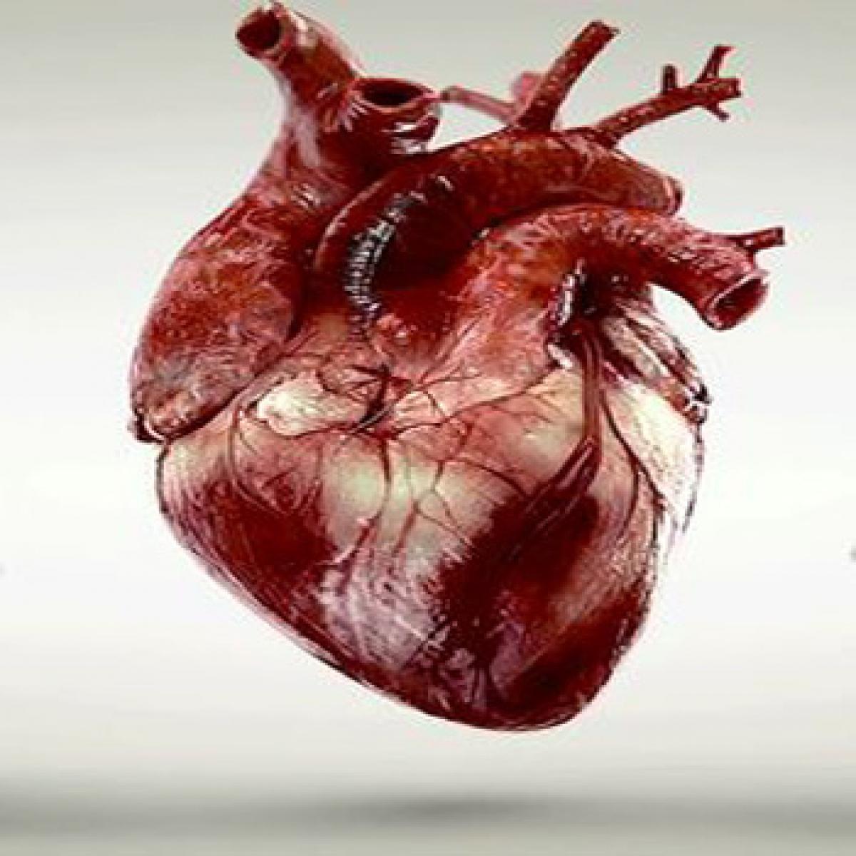
Taking tissue engineering to a whole new level researchers have developed a new technology to build model versions of both heart and liver tissues that function like the real thing. Growing realistic human tissues outside the body, the \"person-on-a-chip\" technology, called AngioChip, could offer a powerful platform for discovering and testing new drugs
Toronto: Taking tissue engineering to a whole new level researchers have developed a new technology to build model versions of both heart and liver tissues that function like the real thing. Growing realistic human tissues outside the body, the "person-on-a-chip" technology, called AngioChip, could offer a powerful platform for discovering and testing new drugs, and researchers believe that the engineered tissues could eventually be used to repair or replace damaged organs.
"It is a fully three-dimensional structure complete with internal blood vessels," said one of the researchers Milica Radisic, professor at the University of Toronto in Canada. "It behaves just like vasculature, and around it there is a lattice for other cells to attach and grow," Radisic noted. The work was published in the journal Nature Materials.
Out of POMaC, a polymer that is both biodegradable and biocompatible, the researchers built a scaffold for individual cells to grow. The scaffold is built out of a series of thin layers, stamped with a pattern of channels that are each about 50 to 100 micrometres wide.
The layers, which resemble the computer microchips, are then stacked into a 3D structure of synthetic blood vessels. As each layer is added, UV (ultraviolet) light is used to cross-link the polymer and bond it to the layer below. When the structure is finished, it is bathed in a liquid containing living cells. The cells quickly attach to the inside and outside of the channels and begin growing just as they would in the human body.
"Our liver actually produced urea and metabolized drugs," Radisic pointed out. The researchers believe that AngioChip could enable drug companies to detect dangerous side effects and interactions between organ compartments long before their products reach the market, saving countless lives.
In future, Radisic envisions her lab-grown tissues being implanted into the body to repair organs damaged by disease. "It really is multifunctional, and solves many problems in the tissue engineering space," Radisic said. "It's truly next generation," she noted.

© 2024 Hyderabad Media House Limited/The Hans India. All rights reserved. Powered by hocalwire.com


