Live
- Farmers demand end to Janjhavathi dispute
- SCR bags 2 All-India performance efficiency shields
- Two persons admitted to hospital as gas leaked at Pharma City
- Mahila Shakti scheme: CS directs officials to set up solar plants in 231 acres
- Remarks against Ambedkar: PCC to take out protest march from Tank Bund to Collectorate today
- TG govt eliciting legal opinion on local quota in med PG admission
- More co-working facilities needed: Sridhar Babu
- State allocates Rs 12.3 cr for CM’s upcoming Davos visit
- AP projects are on accelerated mode: Dinakar
- Rich tributes paid to PV on his death anniv
Just In

Now, your dentist may be able to detect vitamin D deficiency before it becomes a major problem, according to a recent study.
Washington D.C. : Now, your dentist may be able to detect vitamin D deficiency before it becomes a major problem, according to a recent study.
Vitamin D deficiency, a serious but often hidden condition that can now be identified by a simple dental X-ray, McMaster anthropologists Lori D'Ortenzio and Megan Brickley have found.
The researchers and their colleagues had previously discovered that human teeth hold a detailed and permanent record of serious Vitamin D deficiency, or rickets. That record takes the form of microscopic deformities in dentin, the material that makes up the mass of the tooth, and can be extremely valuable for understanding precisely when people, even those who lived centuries ago, were deprived of sunlight, the main source of Vitamin D.
The record is preserved by enamel, which protects teeth from breaking down, unlike bones, which are subject to decay.
The problem with looking for such deformities is that a tooth must be cut open to read the patterns that form a lifetime's Vitamin D record, and the supply of post-mortem teeth available for study is limited.
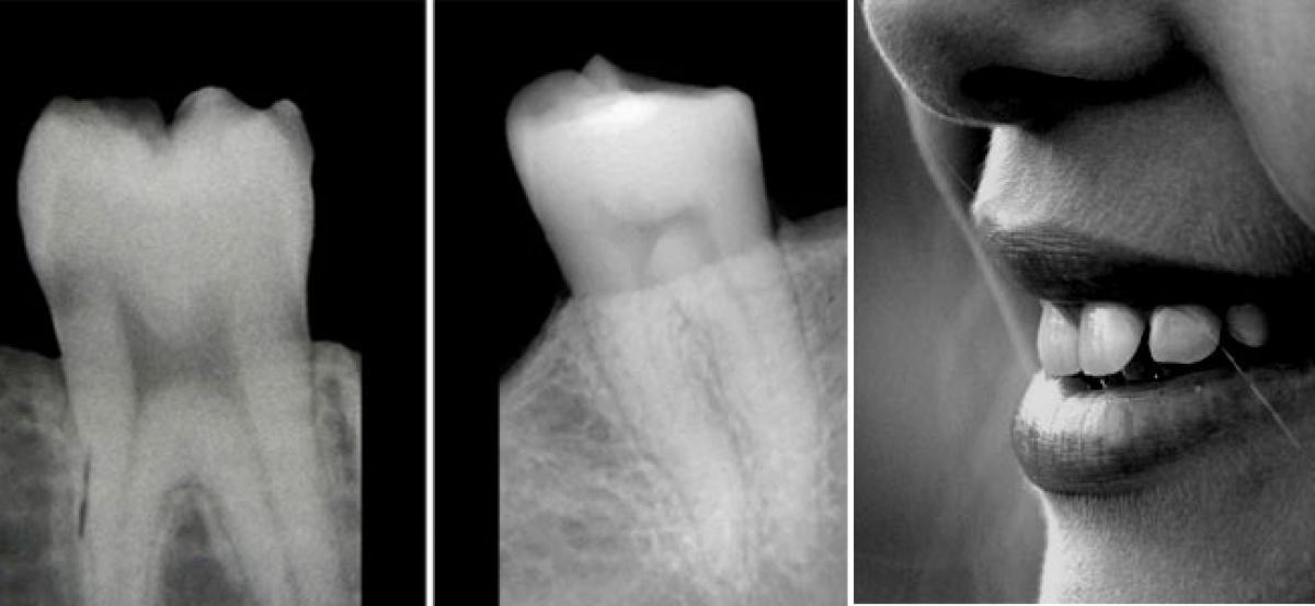
To avoid wasting precious specimens, the researchers went looking for a way to isolate teeth for further study. By using X-rays to study the readily observable shapes of the "pulp horns" -- the dark shadow at the centre of the image of a tooth -- they found a consistent, recognizable pattern that could prove helpful not only to their studies of archaeological teeth, but to living people who may not realize they are suffering from Vitamin D deficiency.
The pulp shape in a healthy person's tooth resembles an arch topped by two cat ears, while the pulp shape of a person who has had a severe deficiency of Vitamin D is asymmetrical and constricted, and typically looks like the profile of a hard-backed chair.
Since the consequences of Vitamin D deficiency can be severe - especially in terms of bone health - knowing who has had a deficiency can help identify people who may have ongoing issues in time to prevent worse damage, the researchers say. If regular dental X-rays show a problem, blood tests can confirm whether there is an ongoing deficiency.
Such evidence could be particularly valuable in the case of children whose bones are still growing, the researchers say, and spotting a problem early could head off future problems with Vitamin-D related bone deficiency.
Knowing more about ongoing Vitamin D deficiency can also help to determine what is the best balance between protecting people from harmful UV rays and making sure they get enough sun to maintain a healthy level of the vital nutrient.
Their research appears in the International Journal of Paleopathology.

© 2024 Hyderabad Media House Limited/The Hans India. All rights reserved. Powered by hocalwire.com


