New MIT-developed MRI sensor to peek deep inside brain

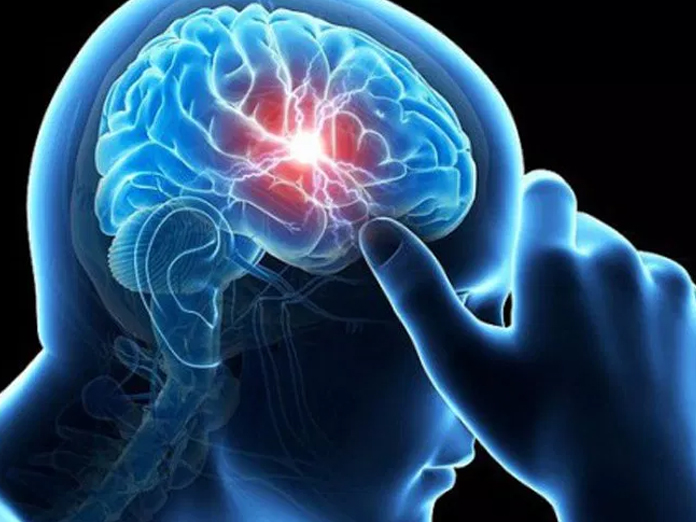
Researchers at the Massachusetts Institute of Technology MIT have devised a new magnetic resonance imaging MRI technique to image calcium activity deep in the brain
New York, Feb 23: Researchers at the Massachusetts Institute of Technology (MIT) have devised a new magnetic resonance imaging (MRI) technique to image calcium activity deep in the brain.
Calcium is a critical signalling molecule for most cells, and it is especially important in neurons.
Using the non-invasive technique, the researchers can track signalling processes inside the neurons of living animals, enabling them to link neural activity with specific behaviours.
However, current imaging techniques can only penetrate a few millimetres into the brain.
"The study describes the first MRI-based detection of intracellular calcium signalling, which is directly analogous to powerful optical approaches used widely in neuroscience but now enables such measurements to be performed in vivo in deep tissue," said Alan Jasanoff, Professor at MIT.
The new MRI sensor, described in the journal Nature Communications, can measure extracellular calcium concentrations.
The team tested their sensor in rats by injecting it into the striatum, a region deep within the brain that is involved in planning movement and learning new behaviours.
They then used potassium ions to stimulate electrical activity in neurons of the striatum, and were able to measure the calcium response in those cells.
The researchers hope to use this technique to identify small clusters of neurons that are involved in specific behaviours or actions.
Because this method directly measures signalling within cells, it can offer much more precise information about the location and timing of neuron activity than traditional functional MRI (fMRI), which measures blood flow in the brain.
"This could be useful for figuring out how different structures in the brain work together to process stimuli or coordinate behaviour," Jasanoff noted.
In addition, this technique could be used to image calcium as it performs many other roles, such as facilitating the activation of immune cells.
With further modification, it could also one day be used to perform diagnostic imaging of the brain or other organs whose functions rely on calcium, such as the heart, Jasanoff said.


